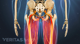


Spondylolysis is a condition in which there is a defect in a portion of the spine called the pars interarticularis (a small segment of bone joining the facet joints in the back of the spine). With the condition of spondylolisthesis, the pars interarticularis defect can be on one side of the spine only (unilateral) or both sides (bilateral). The most common level it is found is at L5-S1, although spondylolisthesis can occur at L4-L5 and rarely at a higher level.
Watch: Isthmic Spondylolisthesis Video
Spondylolysis is the most common cause of isthmic spondylolisthesis, in which one vertebral body is slipped forward over another. Isthmic spondylolisthesis is the most common cause of back pain in adolescents; however, most adolescents with spondylolisthesis do not actually experience any symptoms or pain. Cases of either neurological deficits or paralysis are exceedingly rare, and for the most part it is not a dangerous condition. The most common symptom is back and/or leg pain that limits a patient's activity level.
Since spondylolysis is the most common cause of spondylolisthesis, it may be referred to as an isthmic spondylolisthesis and sometimes these terms are used interchangeably, although this is not correct. There are at least 6 recognized causes of slippage as seen in spondylolisthesis in the literature. According to Dr. Leon Wiltse, these causes are listed as:
- Dysplastic spondylolisthesis (which includes congenital)
- Isthmic spondylolisthesis (which includes lytic or stress fracture, an elongated but intact pars or an acute fracture of the pars)
- Degenerative spondylolisthesis (Pseudospondylolisthesis) — secondary to long-standing degenerative arthrosis (degenerative disc disease and degeneration of the facet joints)
- Traumatic spondylolisthesis (secondary to a fracture of the neural arch)
- Pathologic spondylolisthesis (from bone disease such as metastatic disease, tumor, osteoporosis, etc.)
Importantly, spondylolysis only refers to the separation of the pars interarticularis (a small bony arch in the back of the spine between the facet joints), whereas spondylolisthesis refers to anterior slippage of one vertebra over another (in the front of the spine). Therefore, although the terms are sometimes used interchangeably, this is incorrect and the two are technically not interchangeable.
Watch: Degenerative Spondylolisthesis Video
Watch: Isthmic Spondylolisthesis Video
The underlying cause of spondylolysis has not been firmly established. According to major researchers in spine medicine (including Wiltse, Yochum and Rowe) there have been no recorded cases of spondylolisthesis in a newborn and therefore the condition is not believed to be genetic. Some physicians believe that repetitive trauma (such as from certain sports) may either cause or contribute to the development of spondylolysis.
Spondylolysis or Isthmic Spondylolisthesis Activity Restrictions
In the past, patients have often been advised to limit their activities (especially participation in sports and active exercise) to avoid causing advancement of the spondylolysis. However, new information developed from modern imaging tests and recent research indicates that reduced activity and/or rest to protect the spondylolysis from slipping may not always be necessary. Rest is only necessary if the patient becomes symptomatic. Rest can help eliminate the pain, and when the pain resolves the patient can resume his or her normal activities.
Often adolescents are pulled from their sports participation because of fears that their spondylolysis will lead to spondylolisthesis (slippage of the affected vertebra) and that the slippage will become so severe as to cause permanent damage or paralysis. Adults with spondylolysis are also often counseled to avoid rigorous exercise and/or physically demanding jobs. However, in published medical literature, there are no instances of a patient in a work, industrial, or sports-related environment that has experienced trauma causing spondylolisthesis to slip further and produce neurological deficit or paralysis.
Sophisticated imaging modalities such as single-photon emission computed tomography (SPECT) bone scans and magnetic resonance imaging (MRI) scans of the spine now provide the ability to evaluate the physiological changes that are associated with spondylolysis. This information allows for the important distinction between active and inactive spondylolysis.
- Active spondylolysis. On the SPECT scan an active spondylolysis shows uptake, and an MRI scan shows bone marrow edema adjacent to the pars defect. These findings indicate that there is activity/movement associated with the pars defect, which is likely to produce symptoms of low back pain.
- Inactive spondylolysis. If there are no indications of activity with the pars defect, then the spondylolysis is considered inactive and any low back pain the patient is experiencing is probably incidental (meaning that there is probably another cause of the patient's lower back pain, such as a muscle strain).
Even though activity restriction is not always necessary, careful management of spondylolysis is always advisable. Acute (active) spondylolysis requires more intensive management, while symptoms from spondylolysis that has moved into a chronic (inactive) phase can be managed conservatively.1
References
- 1.Bergmann TF, Hyde TE, Yochum TR. Active or Inactive Spondylolysis and/or Spondylolisthesis: What's the Real Cause of Back Pain? Journal of the Neuromusculoskeletal System. 2002:10:70-78.